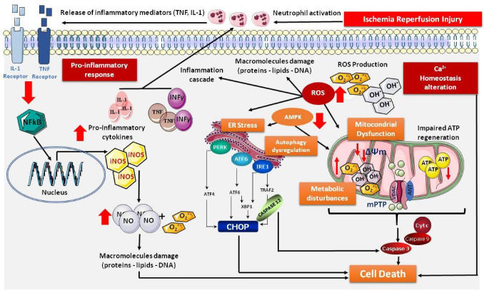
Figure 1.
Schematic representation of the signaling pathways affected by IRI during organ preservation. ↓: decrease; ↑: increase; AMPK: AMP-activated protein kinase; ANT: adenine nucleotide translocase; ATF: activating transcription factor; ATG7: autophagy-related gene 7; ATP: adenosine triphosphate; Ca2+: calcium ion; CHOP: C/EBP homologous protein; CytC: cytrochrome C; ER: endoplasmic reticulum; FKBP: FK506-binding protein; INFγ: interferon-gamma; iNOS: inducible nitric oxide synthase; IL1: interleukin-1; IRE1: inositol-requiring enzyme 1; LC3B: light chain 3 B; mPTP: mitochondrial permeability transition pore; NF-AT: nuclear factor of activated T-cells; NF-κB: nuclear factor kappa-light-chain-enhancer of activated B cells; NO: nitric oxide; O2°− ion: superoxide ion; OH-: hydroxide ion; PERK: protein kinase RNA-like ER kinase; ROS: reactive oxygen species; TNF: tumor necrosis factor; VDAC: voltage-dependent anionic channel.

