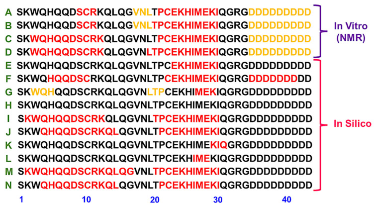
Figure 2.
Lunasin secondary structure content location from the published literature. (A,B) Secondary structure elements identified by NMR for the recombinant lunasin at pH 3.5 without or with disulfide bond, respectively [36]. (C,D) Secondary structure elements identified by NMR for the recombinant lunasin at pH 6.5 without or with disulfide bond, respectively [36]. (E) Proposed α-helix motif with similarity to chromatin-binding proteins [14]; (F) Structural content observed by molecular dynamics simulations [37]; (G,H) Reduced and oxidized forms of the extended lunasin model analyzed by molecular dynamic studies in water [24], respectively. (I,J) Reduced and oxidized forms from the predicted lunasin model analyzed by molecular dynamic studies in water [24], respectively. (K,L) Reduced and oxidized forms, respectively, from the extended lunasin model analyzed by molecular dynamic studies in mixture of water and TFE [24]. (M,N) Reduced and oxidized forms, respectively, from the predicted lunasin model analyzed by molecular dynamic studies in mixture of water and TFE [24]. Residues in red are α-helix motifs and residues in yellow are from a β-strand.

