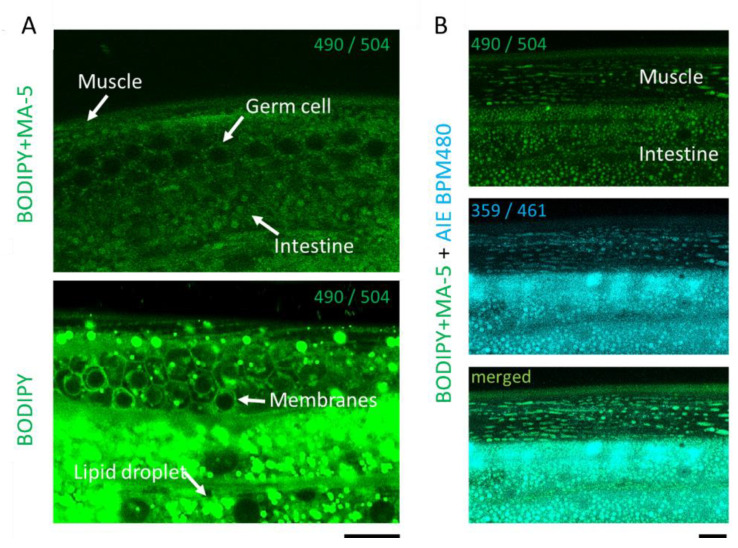
Figure 1.
Penetration and homing activity of MA-5 into intact C. elegans mitochondria. (A) Differences between BODIPY-MA-5 and BODIPY. Fluorescent signals of BODIPY-MA-5 on mitochondria are indicated in muscle, germ cell and intestine (white arrows). BODIPY staining signals show in intestinal lipid droplets and germ cell membranes. (B) The z-stack images of body wall muscle cells of wild-type (N2) adults on day 2 were monitored by confocal microscopy. Localization of MA-5-BODIPY in mitochondria (indicated as green), Mitochondrial Maker AIE mitochondria blue (indicated as blue) and merged image demonstrating mitochondrial localization (indicated as cyan). Fluorescent excitation and emission wavelengths (nm) are indicated as numbers in each picture excitation⁄ emission (nm). Scale bars represent 10 µm.

