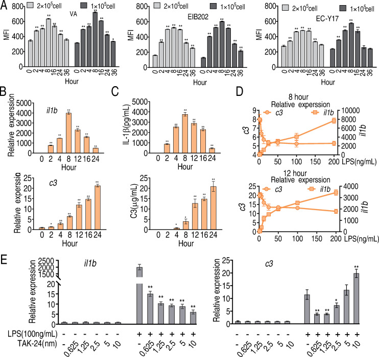
Fig 1. Phagocytosis, IL-1β and C3 in LPS-stimulated macrophages.
A, Phagocytosis of FITC-labeled V. alginolyticus (VA), E. tarda (EIB202), or E. coli (Y17) by LPS-treated RAW264.7-asc cells. Cells were analyzed by flow cytometry and phagocytosis was estimated from mean fluorescence intensity at the indicated time points after LPS-stimulation. Three biological replicates were performed at each time point. B, Transcription of il1b and c3 was estimated by qRT-PCR at the indicated time points after LPS stimulation. C, IL-1β and C3 were quantified in extracellular media by ELISA at the indicated time points after addition of LPS. D, Transcripts of il1b and c3 were quantified by qRT-PCR in cells exposed for 8 or 12 h to the indicated concentration of LPS. E, Transcripts of il1b and c3 were quantified in cells treated with 100 μg LPS. TLR4 Inhibitor TAK-24 at the indicated concentration, or LPS plus TLR4 inhibitor. Results (B-E) are displayed as mean ± SEM, and significant differences are identified (*p < 0.05, **p < 0.01) as determined by non-parametric Kruskal-Wallis one-way analysis with Dunn multiple comparison post hoc test.

