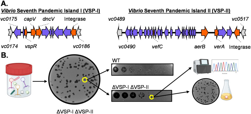
Fig 1. Isolation of VSP-sensitive phages from cholera patient stool samples.
(A) Genomic organization of VSP-I &VSP-II, drawn to scale. Genes characterized before this study are marked in orange. Genes on either island with unknown function are blue. Non-VSP genes are grey. (B) Workflow for isolation of VSP sensitive phage. Filter sterilized supernatant from stool samples was plated on a lawn of the ΔVSP-I ΔVSP-II mutant of V. cholerae. Plaques were picked, diluted and spot plated on multiple hosts to identify changes in plaquing. Plaques were picked and plaque purified before sequencing and further downstream assays. This figure is created with Biorender.com.

