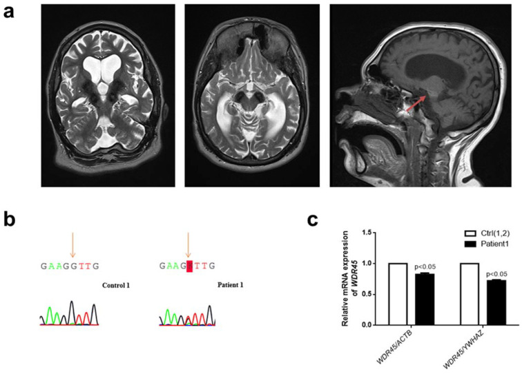
Figure 1.
Clinical and genetic analysis of a novel female patient (Patient 1) with a missense WDR45 variant. (a) Brain MRI from Patient 1. The globus pallidus and the substantia nigra are extensively hypointense bilaterally on axial T2 sequences indicating high levels of iron deposition. The characteristic BPAN T1-hyperintense halo surrounding the substantia nigra is also noted (red arrow). (b) Electropherograms of Sanger sequencing of complementary DNA (cDNA) from cultured fibroblasts from Patient 1 (right panel) and a healthy individual (Control 1) (left panel) in the reverse direction. The variant is highlighted: “K” represents “G” or “T.” (c) The quantification of WDR45 mRNA levels in fibroblasts from Patient 1 compared to fibroblasts from two healthy individuals (Control 1 and Control 2—Ctrl(1,2)). Means and standard error of the mean (SEM) are indicated. Quantification is shown with the mean control level set as 1. The results are based on the mean ratios of WDR45 expression compared to two reference genes, ACTB and YWHAZ. Statistical significance was analyzed by one-way ANOVA (p < 0.05 refers to a significant difference between Patient 1 and two healthy controls (Ctrl(1,2)).

