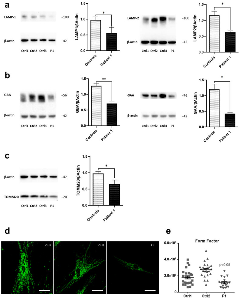
Figure 2.
Disrupted lysosomal and mitochondrial integrity in WDR45-mutant fibroblasts. Western blot analysis of total protein extract from: (a) fibroblasts from WDR45 mutation carrier (P1—Patient 1) and three healthy controls (Ctrl1, Ctrl2, Ctrl3) with antibodies against the lysosomal proteins LAMP-1 (left panel) and LAMP-2 (right panel), and β-actin (loading control), (b) with antibodies against lysosomal enzyme GBA and β-actin (loading control) (left panel) and with antibodies against lysosomal enzyme GAA and β-actin (loading control (right panel). (c) Patient 1’s fibroblasts and three healthy controls with antibodies against the mitochondrial protein TOMM20 and β-actin (loading control). Data analysis was carried out for all Western blots with Ctrl1 set as 1. The error bars indicate standard error of the mean of n ≥ 3 independent experiments. Statistical significance was analyzed by Unpaired t-test (* p < 0.05; ** p < 0.01). (d) The mitochondrial network was visualized by confocal microscopy in fixed cells immuno-stained with anti-GRP75 (green). The scale bar corresponds to 100 μm. (e) A mean form factor was calculated as a measure of mitochondrial interconnectivity by using ImageJ (NIH software). Each dot represents the value from a single cell (15–20 cells per cell culture), and the mean and the error bar (SEM) per individual are indicated. Statistical significance was analyzed by one-way ANOVA (p < 0.05 refers to a significantly decreased form factor in Patient 1’s fibroblasts compared to healthy controls).

