Figure 4.
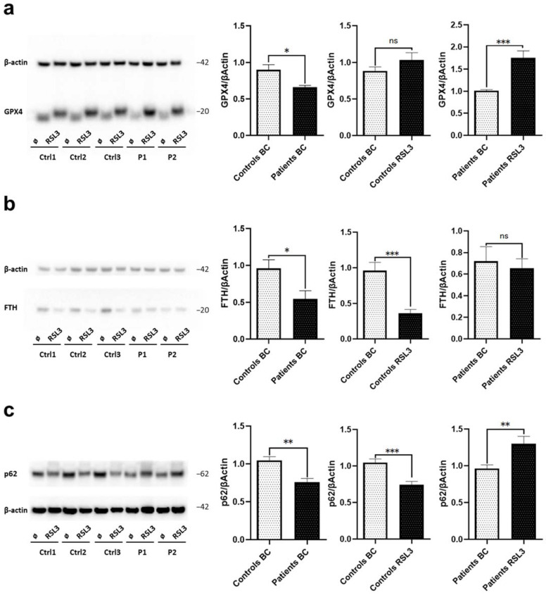
Loss of WDR45 is linked to disrupted iron recycling via ferroptosis. Western blot analysis of total protein extract from fibroblasts from: (a) Patients’ fibroblasts (P1, P2) and three healthy controls (Ctrl1, Ctrl2, Ctrl3) with antibodies against GPX4 and β-actin (loading control) under basal conditions and upon treatment with 1.5 µM of RSL3 for 6 h. (b) WDR45 mutation carriers (Patients, P1 and P2) and three healthy controls (Ctrl1, Ctrl2, Ctrl3) with antibody against FTH and β-actin (loading control) under basal conditions and upon treatment with 1.5 µM of RSL3 for 6 h. (c) Patients’ fibroblasts and three healthy controls with antibodies against p62 and β-actin (loading control) under basal conditions (BC) and upon treatment with 1.5 µM of RSL3 for 6 h. Data analysis was carried out for all Western blots with Ctrl1 in basal conditions set as 1 (first and second diagrams) or P1 in basal conditions (third diagram). The error bars indicate the standard error of the mean of n ≥ 3 independent experiments. Statistical significance was analyzed by Unpaired t-test (ns refers to p ≥ 0.05 (not significant); * p < 0.05; ** p < 0.01; *** p < 0.001).

