Figure 6.
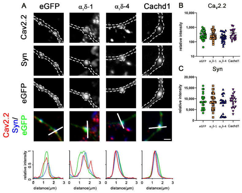
Overexpression of α2δ-1, α2δ-4, or Cachd1 does not increase clustering of N-type channels. (A) Immunofluorescence analysis of axonal varicosities from wildtype neurons overexpressing eGFP and α2δ or Cachd1 constructs, labelled against synapsin and CaV2.2. Micrographs show immunofluorescent signals of CaV2.2 channels at presynaptic boutons, identified by eGFP expression (outlined with a dashed line) and presynaptic synapsin labelling along untransfected dendrites (see also qualitative linescan analysis). Neurons overexpressing α2δ-1, α2δ-4, or Cachd1 exhibited CaV2.2 levels similar to those of the eGFP control. Quantification of CaV2.2 (B) and synapsin 1 (C) shows values for individual cells (dots) and means ± SEM. Cells were obtained from three independent culture preparations. ANOVA with Tukey’s multiple comparison test. (B) 24–39 cells per condition, F(3, 110) = 1.58, p = 0.20. (C) 24–39 cells per condition, F(3, 110) = 0.99, p = 0.40. Scale bar, 1 µm.

