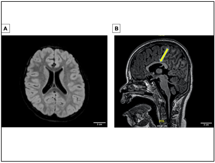
Figure 2.
Brain magnetic resonance image of patient showing multifocal abnormality. Multifocal abnormality with abnormal configuration of lateral and third ventricle, suggestion of minimal infra and supratentorial volume loss, microcephaly, abnormal foliar architecture within the cerebellum, and abnormal appearance of the globes. (A) MRI image of transverse plane showing abnormal lateral ventricles. (B) MRI image of sagittal plane showing abnormal third ventricle indicated by the yellow arrow.

