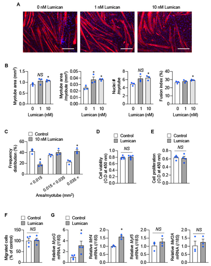
Figure 2.
Lumican-mediated myoblast differentiation. (A–C) C2C12 cells were differentiated with indicated lumican concentrations for 3 days. Myotubes were stained with the anti-myosin heavy chain antibody (A), and morphological parameters of myotubes, such as total myotube area, myotube area per myotube, nuclei # per myotube, and fusion index, were quantified (B). Frequency distribution of myotubes was also determined (C). (D,E) C2C12 cells were treated with or without 10 nM lumican for 24 h, and cell viability (D) and proliferation (E) were examined using Cell counting kit-8 and Cell Proliferation ELISA, respectively. (F) Migration of C2C12 cells was examined using a type IV collagen-coated filter with 10 nM lumican. Migrated cells were quantitated. (G) C2C12 cells were incubated with or without 10 nM lumican for 24 h, and real-time PCR was performed. Each bar represents the mean ± standard error of the mean (SEM). Scale bars: 200 μm. * p < 0.05 vs. the untreated control. NS, not significant.

