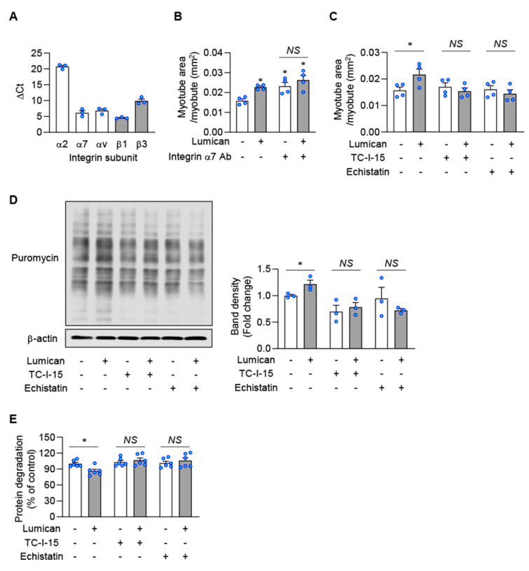
Figure 6.
Lumican-stimulated myogenesis and protein balance mediated by integrins. (A) mRNA levels of integrin subunits in C2C12 cells were determined by real-time PCR. (B) C2C12 cells were differentiated with 10 nM lumican in the presence or absence of integrin α7 neutralizing antibody for 3 days. Myotubes were stained with the anti-myosin heavy chain antibody. Quantitative results of myotube area per myotube are shown. (C) C2C12 cells were differentiated with 10 nM lumican in the presence or absence of integrin inhibitors, such as TC-I-15 and echistatin, for 3 days. Quantitative results of myotube area per myotube are shown. (D) C2C12 cells were pre-treated with or without integrin inhibitors for 30 min. Then, cells were treated with 10 nM lumican for 30 min and underwent lysis after 30 min of incubation with puromycin. Protein synthesis was determined by detecting puromycin-labeled peptides by western blotting. Quantitative results are shown in the right panel. (E) Protein degradation was measured in C2C12 myoblasts cultured with lumican and/or integrin inhibitors, as described in Figure 3. Each bar represents the mean ± standard error of the mean (SEM). * p < 0.05 vs. lumican-untreated control. NS, not significant.

