Figure 2.
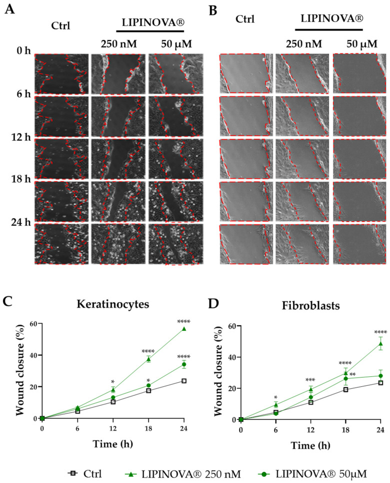
LIPINOVA® treatment accelerates the in vitro migration of keratinocytes and fibroblasts. Representative brightfield images of a scratch assay at different timepoints after scratching a culture plate of keratinocytes (A) and fibroblasts (B) treated with 250 nM or 50 µM of LIPINOVA®, or with saline (Ctrl). Dotted lines define the wound area; scale bar = 100 µm. Quantification of wound closure of keratinocytes (C) and fibroblasts (D) in the presence of LIPINOVA®. Data were normalized to an initial wound area and are represented as mean ± SD percentage. Images were taken at 20× magnification. Experiments were performed in triplicate. Two-way ANOVA was used for statistical analysis. * p < 0.05, ** p < 0.01, *** p < 0.001, and **** p < 0.0001.

