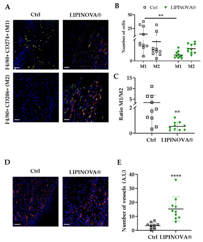
Figure 5.
LIPINOVA® promotes the modulation of Mφ1/Mφ2 phenotype and increases angiogenesis on cutaneous wounds in db/db mice. (A) Representative immunofluorescence images of wound sections stained with specific Mφ1 and Mφ2 antibodies: F4/80+ (red), CD274+ (green), and CD206+ (green). Nuclei are stained with DAPI (blue). (B) Quantification of the M1/M2 macrophage profile ratio. (C) Number of cells stained with antibodies that recognize Mφ1 and Mφ2 in control and LIPINOVA® groups. (D) Representative images of wound sections stained with a caveolin antibody. (E) Quantification of the number of vessels in the wound area; scale bar = 50 µm. Data are represented as the mean ± SD, ** p < 0.01, **** p < 0.0001. n = 10 animals per group.

