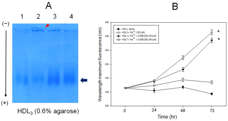
Figure 3.
Proteolytic degradation of HDL by addition of ferrous ions (Fe2+) and enhancement of HDL3 particle stability by the presence of CIGB-258 via the movement of intrinsic Trp fluorescence. (A) Electrophoretic patterns of HDL3 after 72 h incubation with CML and CIGB-258. Lane 1, HDL3 (1 mg/mL) alone; lane 2, HDL3 + Fe2+ (final 120 μM); lane 3, HDL3 + Fe2+ + CIGB-258 (final 20 μM); lane 4, HDL3 + Fe2+ + CIGB-258 (final 40 μM). The red arrowhead indicates the aggregated band by Fe2+ at the loading position. The black arrow indicates the HDL particle band intensity after Coomassie Brilliant Blue staining of apoA-I. (B) Measurement of wavelength maximum fluorescence of the Trp emission scanning spectrum (Ex = 295 nm, Em = 305–400 nm) during 72 h incubation with HDL3 and ferrous ion under the presence of CIGB-258. * p < 0.05 versus HDL3 + Fe2+.

