Figure 2.
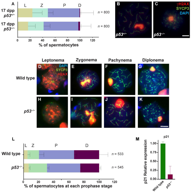
The absence of p53 alters meiotic prophase progression in spermatocytes. (A) Quantification of the percentage of spermatocytes at each prophase stage (leptonema (L), zygonema (Z), pachynema (P), and diplonema (D)) at 17 dpp in p53−/− and p53+/− mice. (n) shows the number of spermatocytes counted per genotype. Bars represent mean ± SD for N = 3 p53+/− mice and N = 3 p53−/− mice; (B) Representative p53+/− pachytene spermatocyte; (C) p53−/− diplotene spermatocytes stained against the axial element protein SYCP3 (green), γH2AX (red), and DAPI (blue); (D–K) Spread chromosomes from representative spermatocytes of p53−/− and wild-type mice from leptonema to diplonema stage, stained against SYCP3 protein (green), γH2AX (red), and DAPI (blue); (L) Quantification of the percentage of spermatocytes at each prophase stage in p53−/− and wild-type adult mice. (n) shows the number of spermatocytes counted per genotype. Bars represent mean ± SD. Scale bar in (C,K) represent 10 µm and apply to all panels; (M) Graph shows relative RNA expression of p21 in wild-type (green) and p53−/− (red) mouse testis by RT-qPCR. Bars represent mean ± SD.

