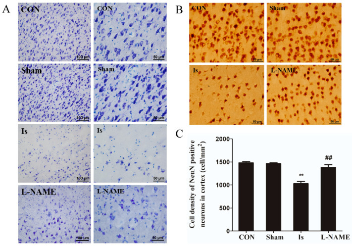
Figure 4.
Effect of cerebral ischemia and L-NAME on cortical neurons of rats (A) Cortical morphology as depicted by Nissl staining, 20× on the left and 40× on the right. (B) Distribution of NeuN-positive neurons in the cerebral cortices by immunohistochemistry staining. (C) Cell density of NeuN-positive neurons in the cerebral cortices. Data are presented as mean ± SEM (n = 6 per group). ** indicates that the difference between the Is and the CON is extremely significant (p < 0.01); ## indicates that the difference between the L-NAME and the Is is extremely significant (p < 0.01).

