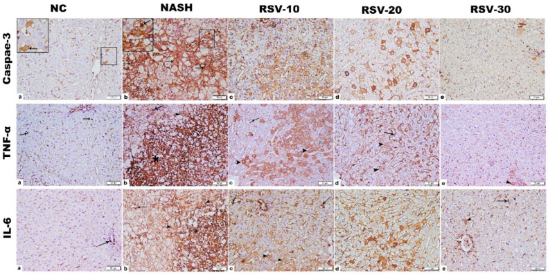
Figure 5.
Photomicrographs of Caspase-3, TNF-α and IL-6 immunohistochemistry-stained liver sections. (1) Caspase-3; (a) NC group: few cells with cytoplasmic positive immunostaining (IS) were detected (inset), NASH group (b): widespread strong cytoplasmic, mainly, and nuclear (inset) positive IS. RSV-10 (c): many cells with cytoplasmic, mainly, and nuclear positive IS. RSV-20 (d): scattered cells with cytoplasmic positive IS. RSV-30 (e) showed few cells with weak cytoplasmic positive IS. (2) TNF-α; NC group (a): cytoplasmic positive immunostaining (IS) in macrophage lining sinusoid (arrow). NASH group (b): widespread strong cytoplasmic positive IS, mainly in the inflammatory cell infiltration (star), in addition to cells lining sinusoids (arrow) and hepatocytes (arrowhead). RSV-10 (c): cytoplasmic positive IS in numerous hepatocytes (arrowhead) in addition to cells lining sinusoids (arrow). RSV-20 (d): cytoplasmic positive IS in scattered hepatocytes (arrowhead) and cells lining sinusoids (arrow). RSV-30 (e): few hepatocytes with weak cytoplasmic positive IS (arrowhead). (3) IL-6; NC group (a): cytoplasmic positive immunostaining (IS) in few cells (arrow). NASH group (b): massive strong cytoplasmic positive IS, mainly in the inflammatory cell infiltration (star), in addition to cells lining sinusoids (arrow) and hepatocytes (arrowhead). RSV-10 (c): cytoplasmic positive IS in numerous hepatocytes (arrowhead) in addition to cells lining sinusoids (arrow). RSV-20 (d): cytoplasmic positive IS in scattered hepatocytes (arrowhead) and cells lining sinusoids (arrow). RSV-30 (e): few hepatocytes (arrowhead) and cells lining sinusoids (arrow) with cytoplasmic positive IS. [Magnification: 200×; inset: 400].

