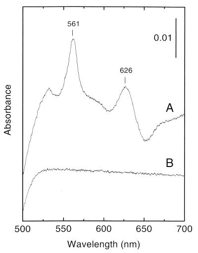
FIG. 1.
Light absorption difference (dithionite-reduced minus air-oxidized) spectra of E. faecalis membranes (3 mg of protein per ml). (A) Membranes from cells grown in the presence of 8 μM hemin; (B) membranes from cells grown in the absence of added hemin. The vertical bar indicates the absorption scale. Difference spectra obtained after oxidation by potassium ferricyanide were identical to those obtained by air oxidation.

