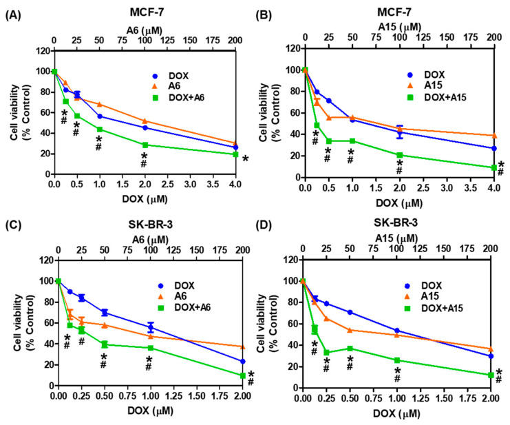
Figure 10.
Cytotoxic effects of A6 or A15 alone or in combination with DOX on MCF-7 and SK-BR-3 cells. (A,B) MCF-7 and (C,D) SK-BR-3 cells were exposed to the indicated concentrations of A6 or A15 alone or in combination with DOX at fixed ratios for 48 h, as illustrated in Figure 9. Control cells were treated with the media containing 0.4% DMSO instead. The IC50 values of A6 and A15 in both cell types, DOX in MCF-7 cells, and DOX in SK-BR-3 cells are set at 50, 1.0, and 0.5 μM, respectively. Cell viability was determined by MTT assay. Data are displayed as mean ± SEM from three independent experiments. # p < 0.05 and * p < 0.05 vs. DOX-treated cells and A6- or A15-treated cells, respectively.

