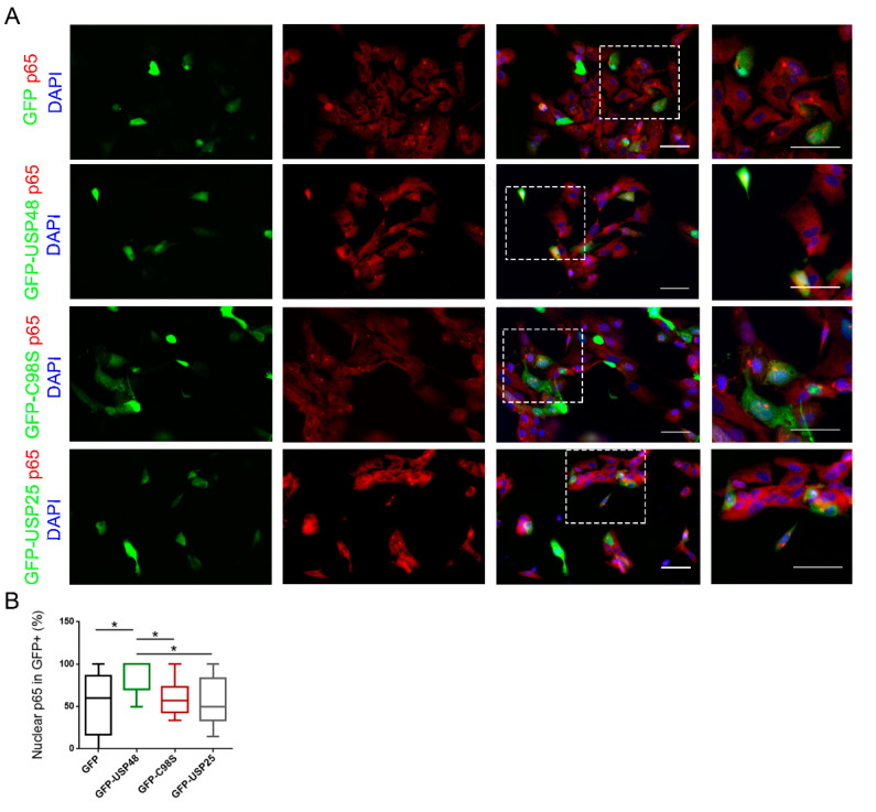
Figure 2.
USP48 overexpression facilitates p65 nuclear localization. (A) Immunofluorescence images from hTERT-RPE1 transfected with either GFP (empty vector), GFP-USP48, GFP-USP48 C98S (catalytically inactive mutant), or unrelated GFP-USP25 (green), and immunostained with p65 (red). Nuclei were counterstained with DAPI (blue). Scale bar: 50 μm in both lower and higher magnification images. (B) Quantification of the percentage of transfected cells presenting nuclear p65. The percentage of GFP-USP48 (WT) expressing cells showing p65 nuclear localization is increased in comparison with either GFP, GFP-USP48 C98S, or GFP-USP25. Box plots (min to max) were obtained from 11–17 images per condition from 2–3 independent replicates. Statistical analysis was performed by one-way ANOVA: * p-value ≥ 0.05.

