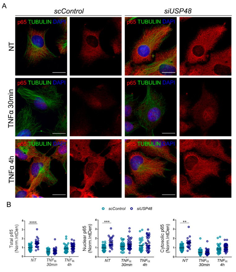
Figure 3.
USP48 downregulation stabilizes p65. (A) Immunocytochemistry of anti-p65 (red) in USP48 knockdown hTERT-RPE1 cells at basal conditions and after treatment with TNFα at two different time points: 30 min (early response) and 4 h (late response). Cells were counterstained with anti-tubulin (green) to highlight the cell shape, and DAPI to delimit the nucleus (blue). (B) Total p65 (Integrated density) was significantly increased in USP48 silenced cells (siUSP48, dark blue dots) in comparison with control cells (scControl, light blue dots), being this increase was detectable in both nuclear and cytosolic compartments. No significant changes were found following TNFα treatment at 30 min or 4 h. Scale bar: 20 μm. Data are represented as the mean, n = 33–45 cells per condition from 3 independent experiments. Statistical analysis by two-way ANOVA: ** p-value ≥ 0.01; *** p-value ≥ 0.001; **** p-value ≥ 0.0001.

