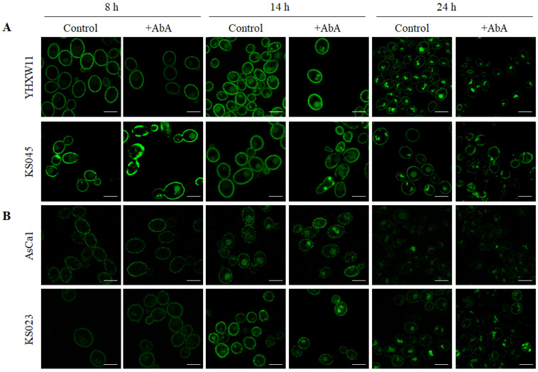
Figure 5.
Subcellular localization of Pma1-GFP (A) and Cdr1-GFP (B) in C. albicans CAF2-1 (YHXW11 and AsCa1) and erg11∆/∆ (KS045 and KS023) strains after 8, 14, and 24 h of culture under control conditions or after treatment with AbA (0.05 μg/mL). Microphotographs were obtain using confocal microscopy. Scale bar 5 μm.

