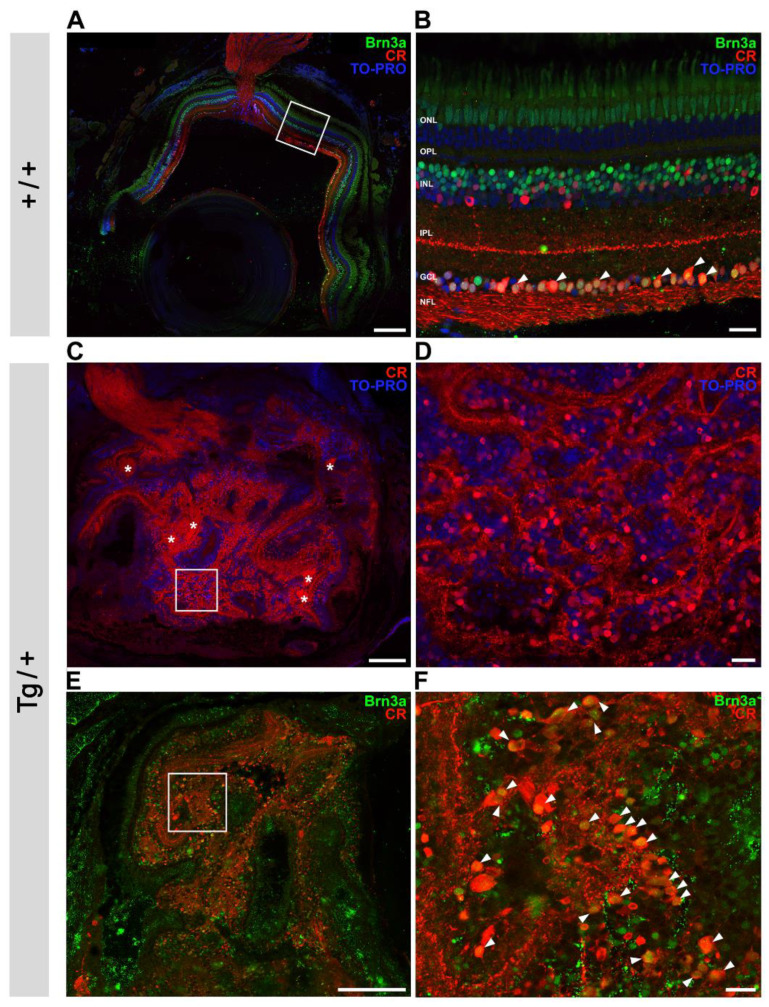
Figure 9.
Increased number of retinal ganglion cells in the retinal mass present in the most severe phenotype of old (two-year-old) myocilin transgenic zebrafish. Overview and detailed images of (A,B) wild-type zebrafish eye and (C–F) myocilin transgenic zebrafish eye. Images in (B,D,F) are details indicated by white boxes in (A,C,E), respectively. (A,B) Double immunostaining for calretinin (CR), a calcium-binding protein, and the transcription factor Brn3a was used for the identification of ganglion cells (arrowheads). Optic nerve fibers were immuno-positive for calretinin. TO-PRO-3-iodide was used for nuclei staining. (C–F) Asterisks: axon bundles. Scale bars in (A,C,E): 200 µm; scale bars in (B,D,F): 20 µm. ONL: outer nuclear layer; OPL: outer plexiform layer; INL: inner nuclear layer; IPL: inner plexiform layer; GCL: ganglion cell layer; NFL: nerve fiber layer; Tg/+: transgenic; +/+: wild type.

