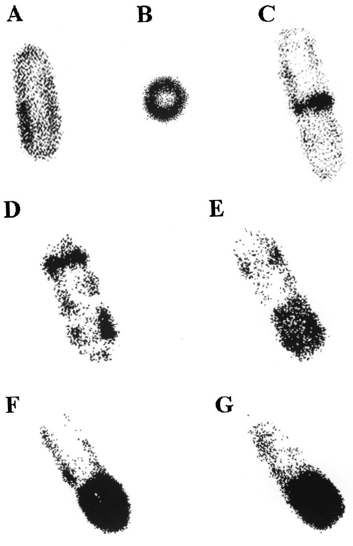
FIG. 2.
FtsH-GFP+ localization in live cells. Cells were grown either in Luria broth (A to C) or in DS medium (22) (D to G) in the presence of 0.1% xylose to induce ftsH-gfp+. FtsH-GFP was visualized in live cells as follows. Cells from 1.5 ml of growth medium were sedimented, resuspended in 200 μl of Tris-HCl buffer (pH 7.4), and mixed with 500 μl of 4% agarose; 10 to 20 μl was applied to a glass slide, covered with a coverglass, and allowed to cool for ∼1 min. A Leica (Heidelberg, Germany) TCS/SP confocal scanning laser microscope equipped with a argon-ion laser for excitation at 488 nm was used. Detection occurred at 510 nm, and the data from the channel were collected with fourfold averaging at a resolution of 512 by 512 pixels and processed using Corel Photo Point 7. Original magnification, ×20,000.

