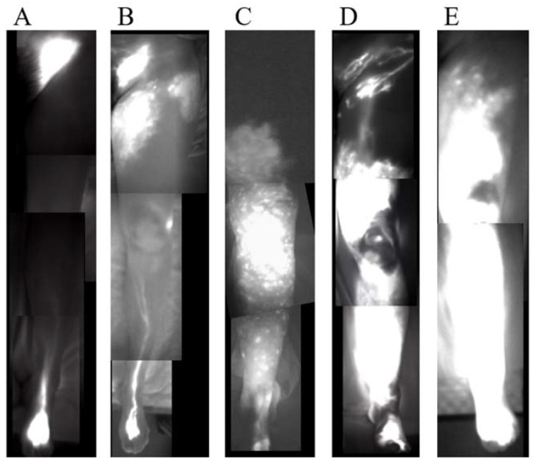
Figure 3.
Indocyanine green (ICG) lymphography images of stage I to V. (A) Stage 1. Splash pattern around the groin region. (B) Stage 2. Stardust pattern extending proximal to the superior border of the patella. (C) Stage 3. Stardust pattern extending distal to the superior border of the patella. (D) Stage 4. The observed stardust pattern extends to the entire limb. (E) Stage 5. The existence of a diffuse pattern with a stardust pattern in the background.

