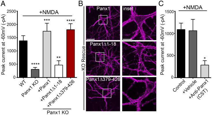
Fig. 7.
Functional importance of Panx1 N terminus. (A) Summary data from whole-cell recordings in neurons from Panx1 KO (n = 19) or WT (n = 28) mice and, additionally, in Panx1 KO neurons expressing Flag-Panx1 (n = 9), Flag-Panx1Δ1–18 (n = 15), or Flag-Panx1Δ379–426 (n = 13). For this set of recordings, NMDA (100 μM) was applied for a duration of 3–5 min. **P < 0.01, ***P < 0.001, and **** P < 0.0001, one-way ANOVA with post hoc Bonferroni test when compared with WT or Panx1 KO. No significant difference was seen between WT, KO+Panx1, and KO+Panx1Δ379–426. (B) Representative images confirming expression and cell surface localization (magenta) of Flag-Panx1, Flag-Panx1Δ1–18, and Flag-Panx1Δ379–426 expressed ectopically in neurons derived from Panx1 KO. Scale bars, 50 µm; inset scale bars, 20 µm. (C) Whole-cell recordings from neurons with control intracellular fluid (ICF) (control) (n = 13) or ICF supplemented with either vehicle (+vehicle) (n = 12) or anti-Panx1 (CST) (+Anti-Panx1 [CST]; 1:400 dilution) (n = 8) applied through patch pipette. For this set of recordings, NMDA was applied for a duration of 3 min. *P < 0.05, one-way ANOVA with post hoc Bonferroni test when anti-Panx1 (CST) was compared with control or vehicle. No significant difference was seen between vehicle and control. Data are represented as mean ± SEM (A and C).

