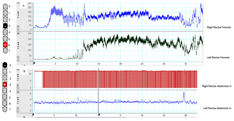
Figure 6.
(A) The captured EMG activities of the right and left rectus femoris m. during standing activity using a standard walker after configuring the right single lead at −3 and +6 and adjusted at a frequency of 20 Hz, 240 µs, and 5–6 volts. (B) The captured EMG activity of the right and left rectus abdominis during sitting at the edge of the mat with use of the upper extremity to perturbate his seated trunk balance after configuring the right lead at −0 and +3 and adjusted at a frequency of 20 Hz, 240 µs, and 5 volts.

