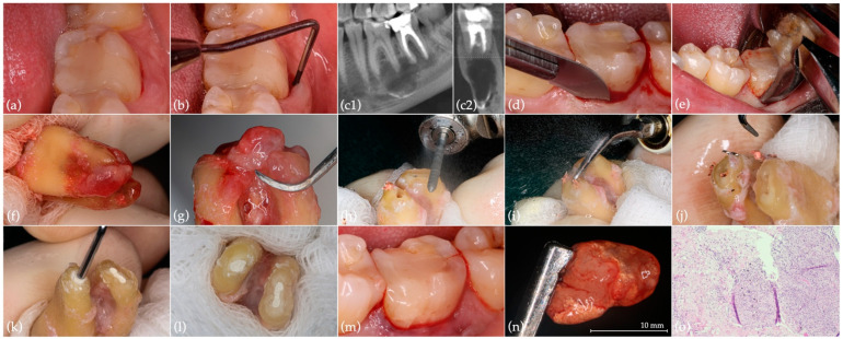
Figure 1.
Preoperative situation and IR of tooth 37: (a) initial situation; (b) mid-buccal probing pocket depth (9 mm); (c1) sagittal and (c2) coronal CBCT section of the region of interest; (d) periodontal fiber separation; (e) atraumatic extraction with dental forceps; (f) meticulous inspection of the root surface; (g) granulation tissue removal using periodontal scalers; (h) root resection; (i) root-end ultrasonic preparation; (j) fractured instrument removal; (k) root-end filling with Biodentine; (l) final aspect of the root-filled apical cavities, with evident isthmus in the mesial root; (m) tooth replanted in the previously prepared alveolus (by a thorough sterile saline solution rinsing and periapical lesion and extruded filling material removal), without splinting; (n) removed large periapical lesion; (o) histological section of the removed periapical lesion showing granulation tissue with abundant inflammatory infiltrate and exogenous material (hematoxylin and eosin staining, 200× magnification).

