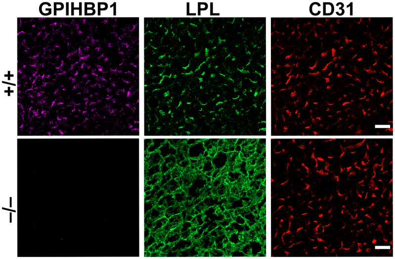
Fig. 1.
Confocal immunofluorescence studies of LPL localization in BAT of Gpihbp1+/+ and Gpihbp1−/− mice. In Gpihbp1+/+ mice (+/+), most of the LPL is located on capillary ECs, bound to GPIHBP1 and colocalizing with CD31 (an EC marker). In Gpihbp1−/− mice (−/−), LPL is mislocalized within the interstitial spaces, bound to HSPGs on the surface of cells (adipocytes and ECs). The LPL in Gpihbp1−/− mice colocalizes with antibodies against collagen IV, a basal lamina protein of adipocytes (17). (Scale bar, 10 μm.)

