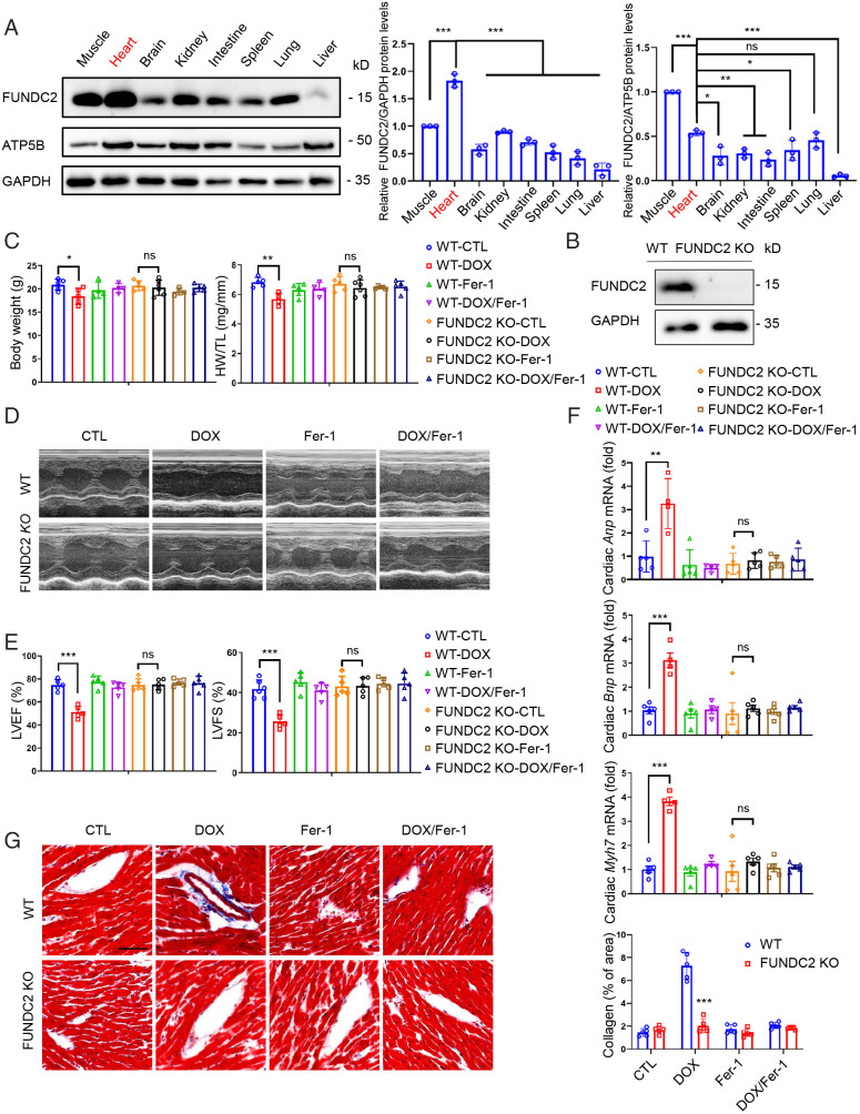
Fig. 1.
Knockout of FUNDC2 ameliorates doxorubicin-induced cardiomyopathy. (A) Relative expression levels of FUNDC2 in different tissues of the male mouse were analyzed by Western blotting (Left) and quantified with GAPDH (Middle) or mitochondrial protein ATP5B (Right) as the internal control (n = 3 mice). (B) Immunoblot of FUNDC2 and GAPDH in heart tissue lysates from WT and FUNDC2-KO male mice. (C–G) Body weight (Left) and heart weight (HW)/tibial length (TL) ratio were measured in WT and FUNDC2-KO mice subjected to DOX or saline (control) in the presence or absence of ferrostatin-1 (Fer-1) at day 4 (n = 4 to 5 mice) (C). For each group subject, the echocardiogram was performed (D); LVEF (Left) and LVFS (Right) were measured (n = 4 to 5 mice) (E); the relative mRNA levels of Anp, Bnp, and Myh7, cardiac hypertrophy biomarkers, were analyzed by qPCR (n = 4 to 5 mice) (F); and the collagen fibrosis in myocardium were visualized by Masson’s Trichrome staining (Left) and quantified by collagen volume fraction (%, collagen fibrosis per total myocardium) (Right). (Scale bar, 50 μm.) (n = 4 to 5 mice) (G). Statistical significance was calculated by Student t test (G) and one-way ANOVA (A, C, E, and F);*P < 0.05, **P < 0.01, ***P < 0.001 and ns indicates no significance.

