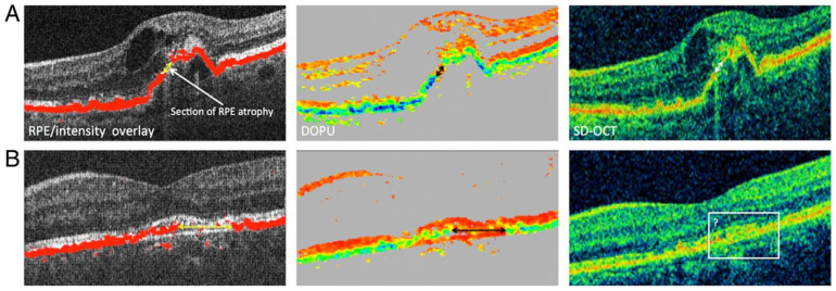
Figure 4.
Polarization-sensitive optical coherence tomography (PS-OCT). (A) Three-figure panel showcases a comparison of PS-OCT (top middle) and SD-OCT (top right) where both modalities are able to identify retinal pigment epithelium atrophy. (B) Three-figure panel showcases a comparison of PS-OCT (bottom middle) and SD-OCT (bottom right) where PS-OCT can more clearly identify the retinal pigment epithelium atrophy. Reprinted with permission from Schütze, C et al. Polarisation-sensitive OCT is useful for evaluating retinal pigment epithelial lesions in patients with neovascular AMD. British Journal of Ophthalmology 2016; 100: 371–377 with license permissions obtained from Creative Commons; Attribution-NonCommercial 4.0 International (CC BY-NC 4.0, https://creativecommons.org/licenses/by-nc/4.0/legalcode (accessed on 1 August 2022)).

