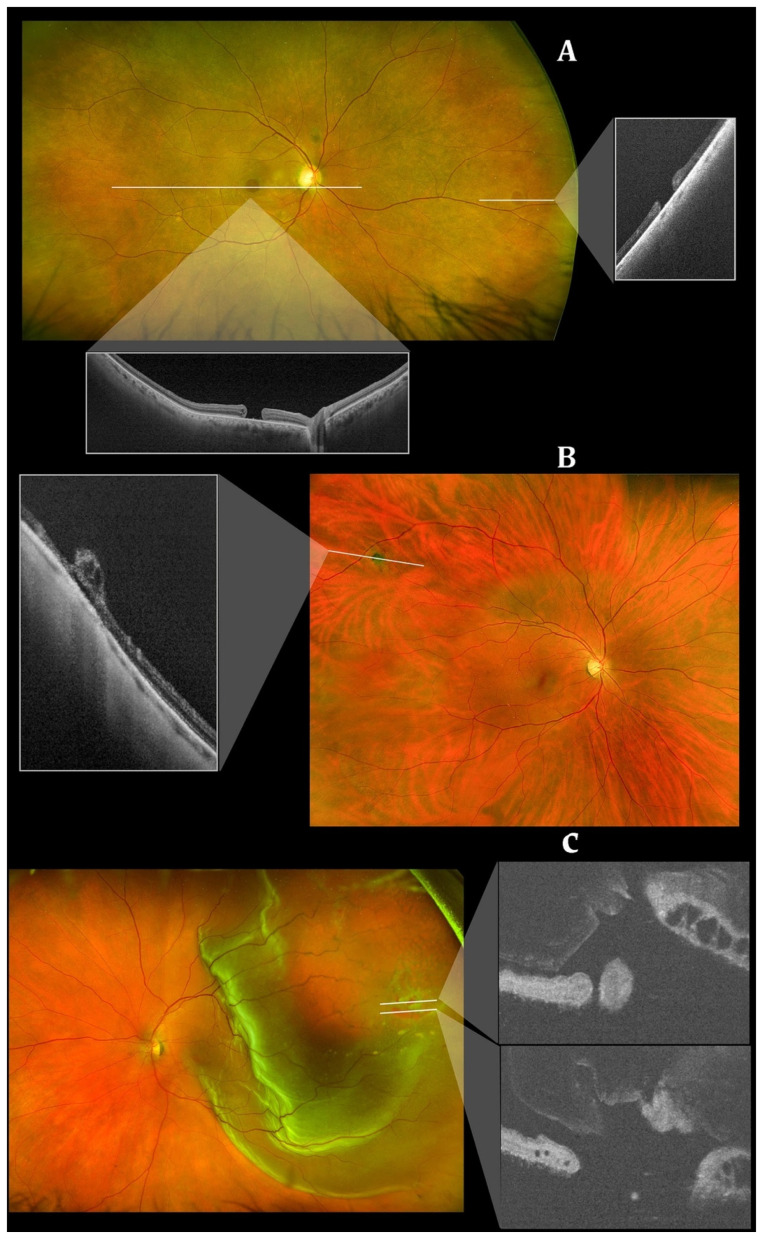
Figure 7.
Integrated scanning laser ophthalmoscope and ultra-widefield imaging for peripheral optical coherence tomography with Optos’ Silverstone swept-source optical coherence tomography (Optos PLC, Dunfermline, UK). (A) A peripheral atrophic retinal hole (right rectangle) and macular hole (lower rectangle). (B) A cystic retinal tuft in the peripheral retina. (C) A retinal detachment in the peripheral retina. Reprinted with permission from Sodhi et al. [96]. Feasibility of peripheral OCT imaging using a novel integrated SLO ultra-widefield imaging swept-source OCT device. Int Ophthalmol 2021; 41(8): 2805-15 with license permissions obtained from Creative Commons; Creative Commons Attribution 4.0 International License (CC BY 4.0, https://creativecommons.org/licenses/by/4.0/legalcode (accessed on 1 August 2022)).

