Figure 1.
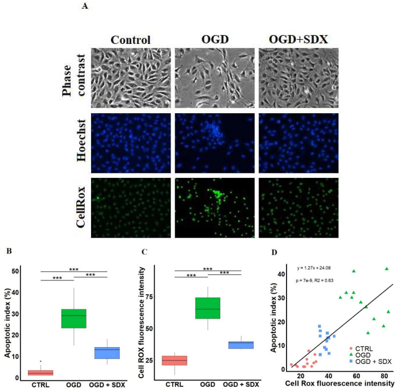
SDX cytoprotective effects against oxidative damage induced by OGD in human endothelial cells. HUVECs were treated for 6 h in OGD in the absence or presence of SDX (0.5 LRU/mL). (A) Microscopic observations. Morphology was visualized and photographed under an inverted phase contrast microscope (original magnification ×200). Apoptotic cells were identified by Hoechst 33342 staining and intracellular ROS production was observed using CellROX Green Reagent. The images were examined under a fluorescence microscope (original magnification ×200). Control: normal conditions; OGD: cells exposed to simulated ischemia in vitro only; OGD + SDX: cells exposed to simulated ischemia in vitro and treated with SDX. Apoptotic index (B) and quantification of CellROX green fluorescence intensity (C) in each corresponding group (n = 12). Data in panels (B,C) are box-plots representing the median and quartiles with the upper and lower limits. Significant results are marked with asterisks (*** p < 0.001); (D) Correlation between apoptotic index and ROS production. Apoptotic index and ROS generation were determined as described in panel (A). Pearson’s correlation coefficient R2 = 0.63 was calculated from the linear regression analysis between apoptotic index and CellROX green fluorescence intensity. Control group. [·] is the oulier.

