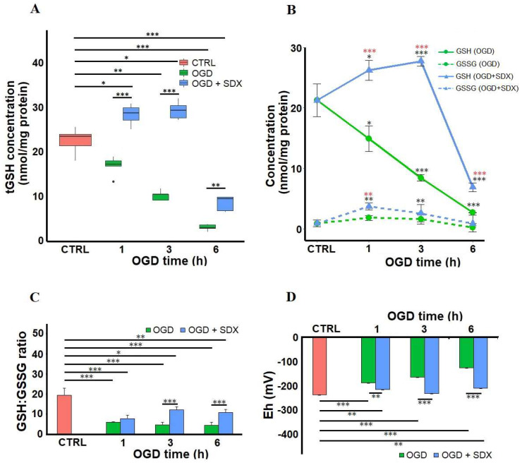
Figure 2.
Time-dependent effects of SDX on the intracellular GSH level and redox state in human endothelial cells subjected to OGD. HUVECs were treated for 1, 3, or 6 h in OGD in the absence or presence of SDX (0.5 LRU/mL). The intracellular concentrations of total glutathione (tGSH) (A), reduced glutathione (GSH), and oxidized glutathione (GSSG) (B) were measured by colorimetric assay (n = 4–6). The tGSH, GSH, and GSSG levels were normalized to total protein concentrations and expressed as nmol/mg protein. The GSH:GSSH ratio (C) and the GSH redox potential (ΔEh) (D) for each incubation period were calculated. CTRL: normal conditions; OGD: cells exposed to simulated ischemia in vitro only; OGD + SDX: cells exposed to simulated ischemia in vitro and treated with SDX. Data in panel (A) are box-plots representing the median and quartiles with the upper and lower limits. Each point in panel (B) and bar graphs in panels (C,D) represent mean ± standard deviation (SD). In panels (A–D), the asterisks indicate the statistically significant differences (* p < 0.05; ** p < 0.01; *** p < 0.001). In panel B, statistically significant differences between OGD groups and OGD + SDX groups are marked with red asterisks (*), while black asterisks (*) are used to mark the differences between the test groups and Control group. [·] is the oulier.

