Figure 4.
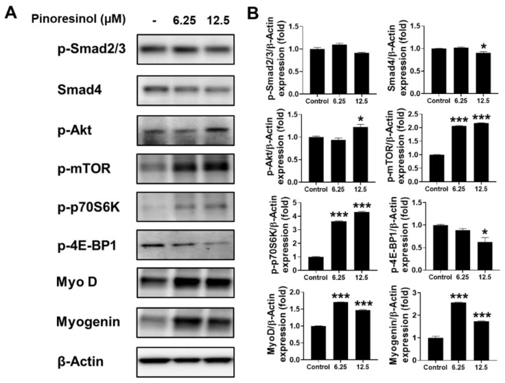
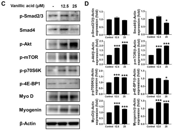
Activated myogenesis by pinoresinol and vanillic acid in mouse myoblast cell line. Western blot using C2C12 cells subjected to different concentrations of pinoresinol (A) or vanillic acid (C) to assess TGF-β signaling (p-Smad2/3 and Smad 4), IGF-1 signaling (p-Akt, p-mTOR, p-p70S6K, and p-4E-BP1), MyoD, and myogenin. β-Actin was used as the loading control. The relative band intensity of proteins to that of β-actin is expressed as the fold change compared to the control (no treatment) for pinoresinol (B) or vanillic acid (D). Data are expressed as mean ± SEM, obtained from at least triplicate determinations. * and *** indicate p < 0.05 and p < 0.001 by one-way ANOVA, respectively.


