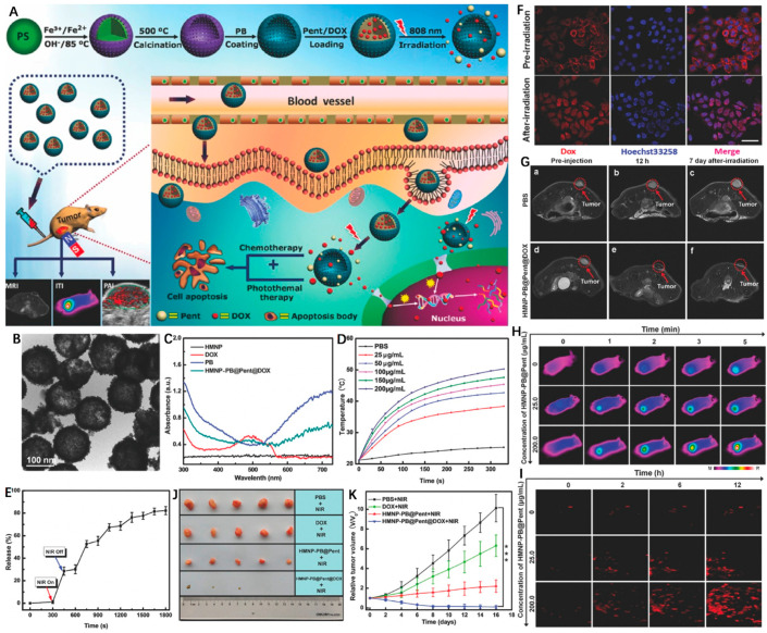
Figure 9.
(A) Schematic illustration of the design and synthesis of HMNP-PB@Pent@DOX for NIR-guided synergetic chemo-photothermal tumor therapy with tri-modal imaging in vivo. (B) TEM image of HMNP-PB. (C) Representative UV–Vis–NIR absorption spectra of HMNP, DOX, PB, and HMNP-PB@Pent@DOX NPs in PBS. (D) Concentration-dependent thermogenesis of HMNP-PB@Pent@DOX in PBS irradiated by NIR laser (808 nm, 1.2 W cm−2, 5 min). (E) The on–off switch drug release of HMNP-PB@Pent@DOX NPs in PBS with NIR laser (808 nm, 1.2 W cm−2). (F) CLSM images of HepG2 cells incubated with HMNP-PB@Pent@DOX NPs before and after irradiation by NIR laser (808 nm, 1.2 W cm−2) for 5 min. (G) T2-MR images of tumors of (a) preinjection of PBS, (b) after injection of PBS for 12 h, (c) after injection of PBS for 7 d with NIR laser irradiation, (d) preinjection of HMNP-PB@Pent@DOX, (e) after injection of HMNP-PB@Pent@DOX for 12 h, and (f) after injection of HMNP-PB@Pent@DOX for 7 d with NIR laser irradiation. (H) Infrared thermographic images of tumor-bearing nude mice irradiated with NIR laser. (I) PAI signals in the tumors before and after injection of HMNP-PB@Pent@DOX for 2, 6, and 12 h. (J) Photographs of the solid tumors after different treatments for 16 d. (K) Relative tumor volumes of mice with different treatments. Reprinted with permission from Ref. [125]. Copyright 2017, Wiley-VCH GmbH. *** p < 0.001.

