Figure 2.
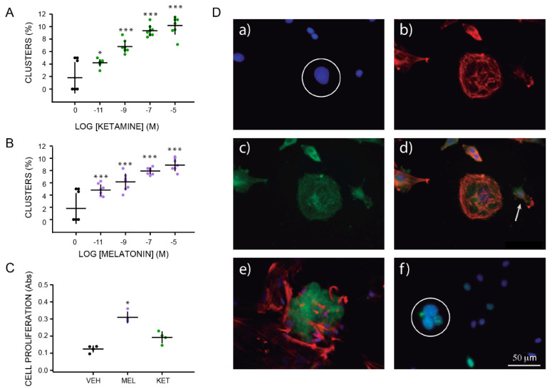
Characterization of clusters formed by olfactory neuronal precursors cultured with either ketamine or melatonin. Graph of the frequency of cluster formation after 6 h of incubation with increasing concentrations of either KET (10−11 to 10−5 M) or MEL (10−11 to 10−5 M) are shown in Panels A, and B, respectively. Formation of clusters was evaluated by counting them in each of 10 chosen fields (n = 10) and normalized by the total number of cells per field determined by DAPI staining. Cell proliferation induced by treatment with vehicle (VEH), MEL (10−7 M), or KET (10−7 M) were measured by the WST1 transformation by mitochondrial enzymes to formazan and shown in Panel C. Each point represents the mean of absorbance values obtained in 4 wells. The mean ± standard deviation of the mean (SEM) is from 1 or 2 experiments performed by quadruplicate. Each experiment was repeated three times. Data were analyzed by one-way ANOVA with Bonferroni’s post-test. * p = 0.009; *** p = 0.001 when compared with vehicle (VEH) control. Cell proliferation was analyzed by Kruskal–Wallis followed by Dunn´s post-test p = 0.001. Panel D shows immunofluorescent labeling of ONPs cultured with KET 10−7 M during 48 h and stained with DAPI (a), phalloidin-TRITC (b), and anti-nestin antibody followed by a secondary antibody coupled to FITC (c). Merged images show colocalization of these labels (orange) in a spherical cluster (d). Image e shows a merged image of a spherical cluster simultaneously labeled with an anti-doublecortin antibody followed by a secondary antibody coupled to FITC (green) and phalloidin-TRITC (red). A representative image of clusters stained with DAPI (blue), pre-labeled with BrdU and stained with a specific anti-BrdU antibody (green) is shown in image f. Spherical clusters are noted by a white circle in images a and f. A single cell is noted in d with an arrow. Bar = 50 µm.

