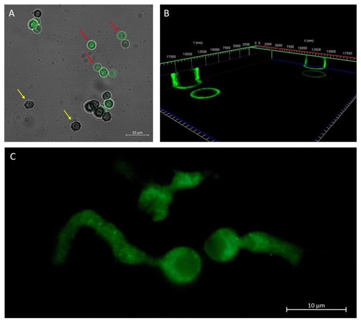
Figure 2.
Overlap of the bright field image and the fluorescence image, which shows A. brasiliensis conidia treated for 10 min with 50 nm PLGA-coumarin6-NPs. In the first stage of conidia development (yellow arrow) the protective envelope did not allow interaction with NPs. In a later stage of conidia development (red arrow), when the envelope broke, fluorescence along the conidia capsule was observed (A). A 3D reconstruction of A. brasiliensis conidia treated with NPs for 10 min. The fluorescence signal was detected along the wall of the conidia (B). Fluorescence image of the hyphae of the newly germinated A. brasiliensis conidium treated with 50 nm PLGA-coumarin6 -NPs. The fluorescence signal inside A. brasiliensis hyphae is visible after 1 h of NPs administration (C).

