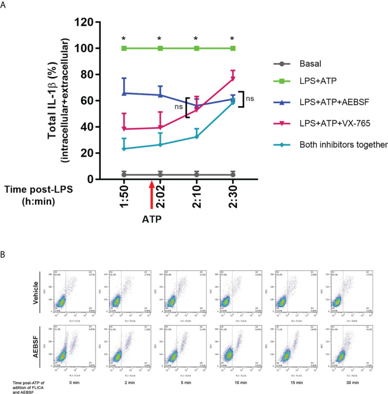
Figure 7.

Dynamic of pro-IL-1β processing by caspase-1 and NSPs. (A) Total (intracellular+extracellular) mature IL-1β levels determined by ELISA in neutrophils stimulated with LPS+ATP either in the presence of AEBSF, VX-765 (caspase-1/4 inhibitor) and both inhibitors together, added at different time points before and after inflammasome activation with ATP. Neutrophils were stimulated or not with LPS and 2 h later with ATP. Ten min before ATP treatment or 2, 10 or 30 min after, cells were treated with AEBSF (0.35 mM), VX-765 (50 µM) or both inhibitors together. At 5 h post-LPS stimulation, total (intracellular+extracellular) concentrations of mature IL-1β were determined by ELISA. Data are depicted as % of the levels of mature IL-1β produced upon LPS+ATP stimulation alone and represent the mean ± SEM of experiments performed in duplicate with 4 donors. *p<0.05 LPS+ATP vs each treatment at the corresponding time point; ns: non-significant; Two-way ANOVA with Tukey’s multiple comparisons test. (B) Effect of inhibition of NSPs at different time points after inflammasome activation with ATP on caspase-1 activity. Neutrophils were stimulated with LPS for 2 h, and then were treated with ATP to induce inflammasome activation. At the time points post ATP-addition indicated below each dot plot, FLICA was added or not simultaneously with AEBSF to trap during a 5 minutes-lapse all active caspase-1. Images depict representative dot plots of experiments performed with 4 donors showing FLICA fluorescence of neutrophils after having excluded the doublets.
