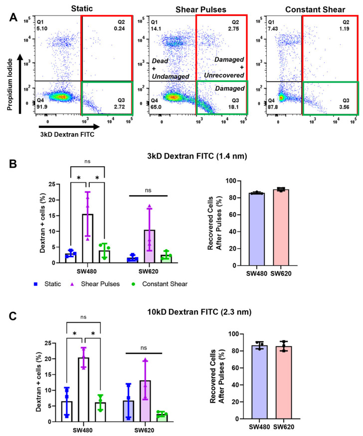
Figure 4.
Shear pulses cause cell-membrane damage, indicated by membrane-pore formation, which is rapidly repaired. (A) Representative flow plots demonstrating enhanced dextran internalization after pulses of FSS. The percentages of damaged cells are shown in red and green (dextran+, Q3 and Q2), while the percentages of damaged cells that were unrecovered are shown in green (dextran+ and PI+, Q2 only). (B,C) Quantification of cell-membrane damage and repair for 3kD and 10kD MW dextran, respectively. Percentage of recovered cells was calculated by dividing the population of damaged and recovered cells (Dextran+/PI+, Q3) by the total number of damaged cells (Dextran+, Q2 + Q3). N = 3, ns = not significant, * p < 0.05 (two-way ANOVA with multiple comparisons). Error bars represent mean ± SD.

