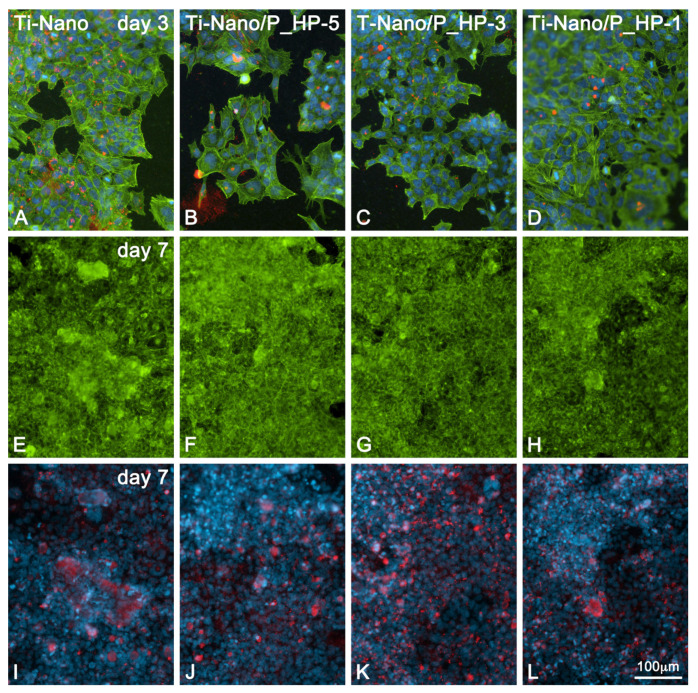
Figure 5.
Epifluorescence of UMR-106 cells grown on Ti-Nano surfaces functionalized with 1 × 10⁻⁵, 1 × 10⁻³, and 1 mg/mL grape-pomace extract and the control surface on days 3 (A–D) and 7 (E–L) of culture. (Ti-Nano, Ti-Nano/P_HP-5, Ti-Nano/P_HP-3, and Ti-Nano/P_HP1) Green, red, and blue fluorescence depicts actin cytoskeleton, bone sialoprotein (BSP), and cell nuclei, respectively. Note the BSP-positive mineralized matrix foci in (I–L). The microscopic fields are the same respectively for (E-I, F-J, G-K, H-L).

