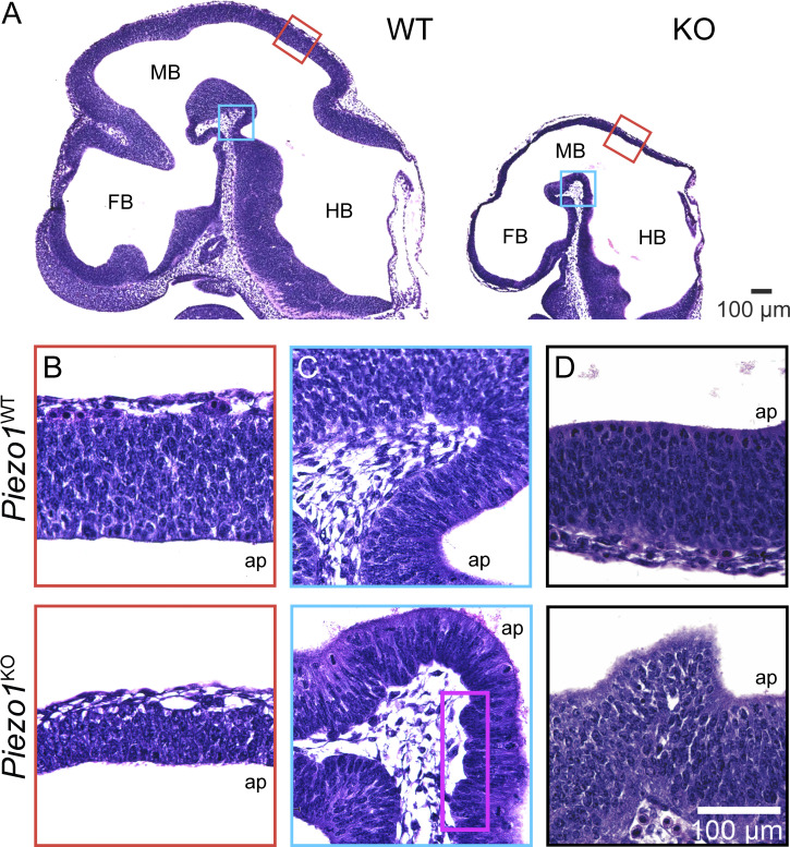
Figure 1.
Histological analysis reveals abnormalities in the developing brain of Piezo1 KO mutant mice. (A) Representative H&E stains of E10.5 WT (left) and Piezo1 KO (right) littermate embryo sections. FB, forebrain; MB, midbrain; HB, hindbrain. Regions marked by red and blue boxes are shown at higher magnification in B and C. (B–D) Representative images highlighting differences in neuroepithelial thickness (B), inner/apical and outer/basal border morphology (C and D), and pseudostratified layering (D) between E10.5 WT (top row) and Piezo1 KO (bottom row) littermates. Purple box in C highlights the abnormal undulations at the basal border in the mutant. Images in A–D are representative of n = 7 embryos for WT and for Piezo1 KO. Scale bar in D also applies to B and C.

