FIGURE 1.

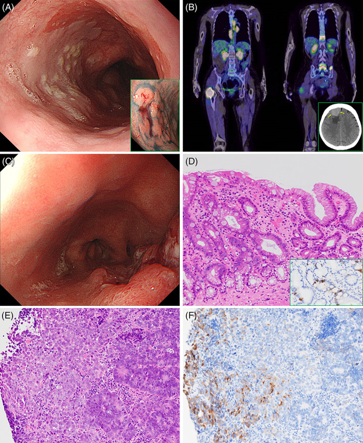
Clinicopathological findings of metachronous carcinomas derived from long‐segment Barrettʼs esophagus (LSBE). (A) LSBE spreading distally to 20 cm from the incisor teeth on upper endoscopy, with type 0‐IIa tumor (tubular adenocarcinoma) at the 23 cm incisor (inset). (B) Positron emission tomography–CT reveals multiple osseous metastases (11th thoracic vertebra, 5th lumbar vertebra and bilateral iliac bones), accompanied by osteolytic masses in the spinous process of the 5th lumbar vertebra and the proximal part of the right femur. Furthermore, left supraclavicular fossa (not included in this image), mediastinal, and paraaortic lymph nodes also show radioactive tracer accumulation. Inset: site of tumor location in the right frontal lobe on a CT slice (yellow arrows). (C) Advanced type 3 cancer, located 35 cm from the incisor, in the LSBE. (D) LSBE histologically possessing foveolar (right side) as well as metaplastic intestinal epithelia (left side) (H&E), with scattered chromogranin A‐immuno‐positive neuroendocrine cells (inset). (E) On biopsy sections, the type 3 esophageal tumor is comprised of an admixture of moderately to poorly differentiated adenocarcinoma (right side) and undifferentiated components (left side) (H&E). (F) Synaptophysin staining immunohistochemically demonstrates the neuroendocrine nature of the pattern‐less neoplastic region (left side)
