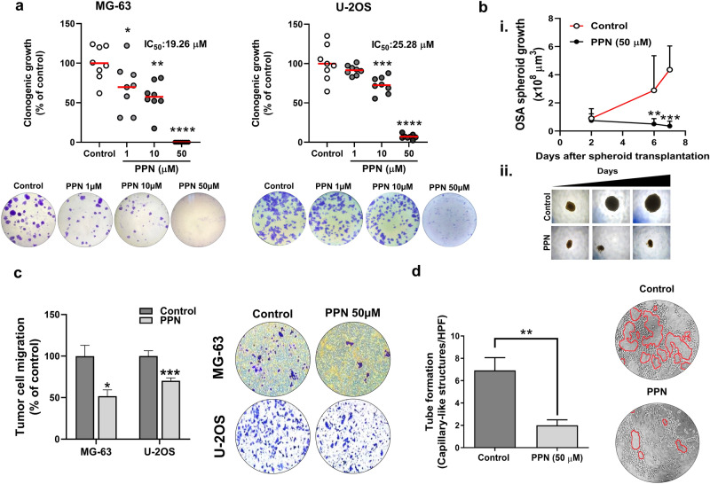
Figure 2.
Propranolol abrogates colony formation, three-dimensional spheroid growth, cell migration and tube formation in osteosarcoma. (a) Number of osteosarcoma (OSA) colonies per well quadrant was quantified after a 7 day incubation with propranolol (PPN, 1–50 µM). Representative microphotographs of MG-63 and U-2OS clonogenic cultures (bottom) in control and PPN 1, 10 and 50 µM groups (left to right) are depicted. (b) PPN completely impairs growth of fully formed OSA spheroids. (i) OSA spheroid volume was assessed as a direct parameter of spheroid growth during the following week. (ii) Representative images of OSA spheroids at days 2, 6 and 7 (left to right). ×40 final magnification. (c) Inhibition of MG-63 and U-2OS cell chemotaxis after a 16 h treatment using PPN at 50 µM. Representative pictures of tumor cells on the bottom of the Transwell inserts (bottom) corresponding to the control or PPN group (left and right, respectively) are shown. (d) Inhibition of capillary-morphogenesis on a Matrigel-coated substrate by MG-63 cells after 16 h treatment with PPN 50 µM. Representative images of capillary-like tubular structures (identified in red lining) in different experimental conditions (×100 magnification). *p < 0.05, **p < 0.01, ***p < 0.001 and ****p < 0.0001. ANOVA followed by Tukey's test for (a), unpaired two-sided t test for (b) and (c) and Mann–Whitney test for (d).

