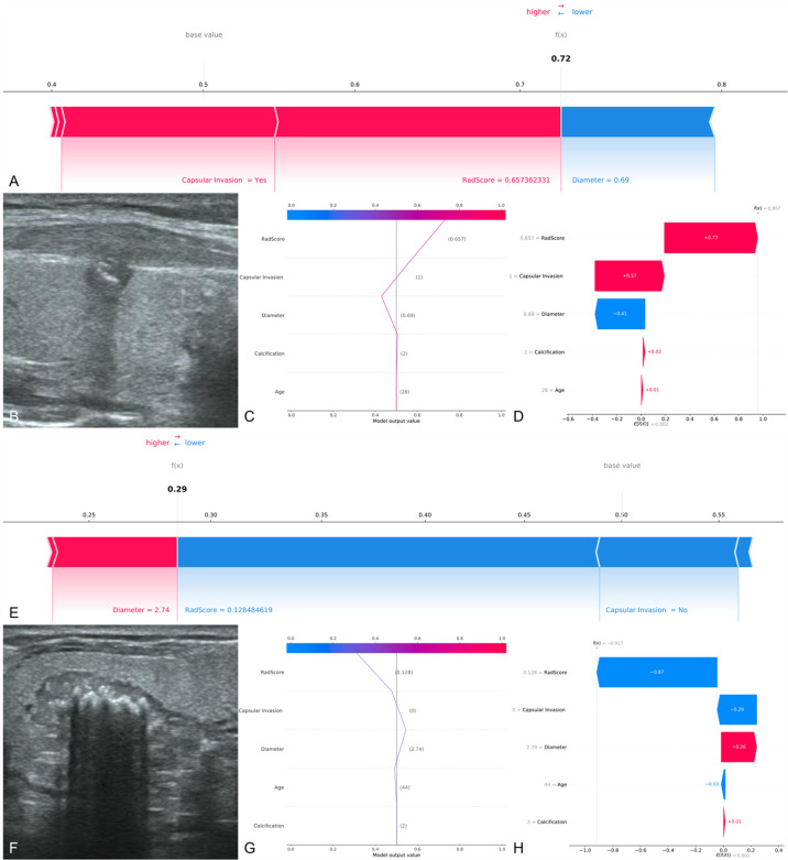
Figure 6.
Two examples of correct prediction of CCLNM+ and CCLNM-. (A–D) A 28-year-old male patient was admitted to our hospital for further treatment after physical examination found a nodule in the right lobe of the thyroid. Ultrasound examination showed a nodule in the middle of the right lobe of the thyroid, 0.69 cm in diameter, with coarse calcification and capsular invasion. Postoperative pathological findings: papillary carcinoma of the right lobe of thyroid, with lymph node metastasis in the right central region. Retrospective analysis of the case showed that the radiomics score was 0.657 and the XGBoost model predicted CCLNM+ correctly. (E–H) A 44-year-old male patient was admitted to our hospital because or volume increase of the right thyroid lobe. Ultrasound examination showed a nodule in the middle of the right lobe of thyroid, 2.74 cm in diameter, with coarse calcification and without capsular invasion. Postoperative pathological findings: papillary carcinoma of the right lobe of thyroid, with no lymph node metastasis in the right central region. Retrospective analysis of the case showed that the 4adiomics score was 0.128 and the XGBoost model predicted CCLNM- correctly.

