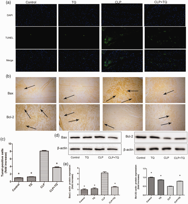
Figure 3.
(a) TUNEL- (green fluorescence) and DAPI-stained (blue fluorescence) photomicrographs. Magnification, ×40. (c) Quantification of apoptotic cardiomyocytes. * P < 0.05 vs. CLP group. (b) Representative immunohistochemical staining for Bax and Bcl-2 in cardiac tissue. Magnification, ×40. Arrows indicate positively stained cells (n = 3). (d) Immunoblotting for Bax and Bcl-2 in cardiac tissue and (e) Bar graph presenting the quantification of Bax and Bcl-2 protein expression. Data are presented as the ± standard error of the mean (n = 3 per group). *P < 0.05 vs. CLP group.
CLP, cecal ligation and puncture; TQ, thymoquinone.

