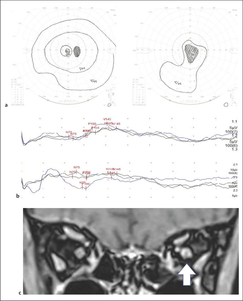
Fig. 2.
Visual field testing by Goldman perimetry (a) has been shown central scotoma on pretreatment. The pretreatment VEP (b) showed higher levels of the waveform and no latencies of P100 are shown for the right eye (upper) and the left eye (lower). Postcontrast orbital magnetic resonance imaging with fat suppression on the coronal image (c) shows the left optic nerve sheath enhancement (indicated by the white arrow).

