Figure 2.
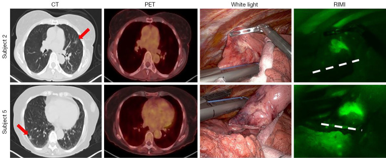
IMI during robotic-assisted thoracic surgery (RIMI) identifies pulmonary GGOs during robotic resection. This figure shows representative images from two patients enrolled in the study. Preoperative CT and PET scans are shown at left, with lesions marked by red arrows. The rightmost two columns show intraoperative white light and near-infrared images, with RIMI clearly identifying both lesions. The dashed line in the rightmost column indicates the stapled resection margin. CT, computed tomography; PET, positron emission tomography; GGOs, ground glass opacities; IMI, intraoperative molecular imaging.

