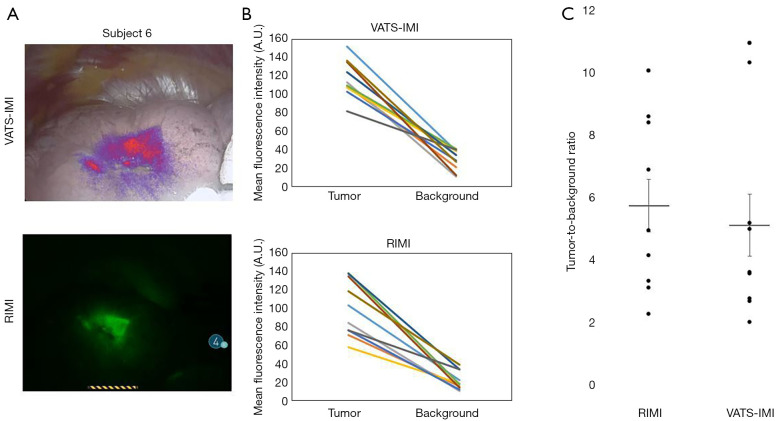
Figure 4.
Comparison of IMI during robotic-assisted thoracic surgery (RIMI) to intraoperative molecular imaging during video-assisted thoracic surgery (VATS-IMI). Panel A depicts representative images comparing VATS-IMI and RIMI in the same patient. Panel B shows mean fluorescence intensity (MFI) of lesion and background measurements, stratified by imaging modality (RIMI vs. VATS-IMI). Each line links lesion and background measurements from the same subject. Panel C depicts tumor-to-background ratio (TBR) of lesions compared to normal lung parenchyma as background, when stratified by imaging modality. Each point on the plot represents a TBR for an individual patient. Mean TBR was >2 for both RIMI and VATS-IMI, and there were no significant differences between groups. A.U., arbitrary unit; IMI, intraoperative molecular imaging.

